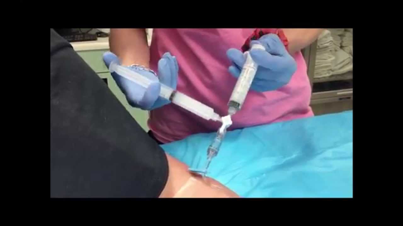
If playback doesnt begin shortly try restarting your device. The bubble test is where they stick an IV in and pump it full of saline that has been shaken as fast as they can.
![]() Transesophageal Echocardiogram Tee Bubble Study Agitated Saline Download Scientific Diagram
Transesophageal Echocardiogram Tee Bubble Study Agitated Saline Download Scientific Diagram
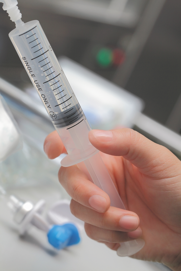
For the bubble study you will get an intravenous IV line in a vein in your arm.
Heart bubble test. Houston Methodist DeBakey Heart. The TEE probe is much closer to the heart since the esophagus and heart are right next to each other. A transesophageal echocardiogram is done by inserting a probe with a transducer down the esophagus.
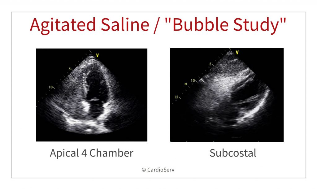
This is sometimes called a bubble study. A bubble study is a noninvasive test that allows physicians to assess the flow of blood through the heart. The bubble study helps to identify those abnormalities.
A bubble echocardiogram is the same procedure as an echocardiogram except an IV is placed in the patients arm. This medical test may be ordered for a patient who appears to be experiencing problems related to the physical function of the heart such as leaky valves or an oversized heart. An EDAC bubble detector sensor was attached prior to the saline injection site and distal to the HeartMate I to measure the size and volume of the bubbles.
This technique was repeated using 05 mL of air and 95 mL of saline bolus and 2 mL of air and 8 mL of saline bolus. A bubble echo involves performing an echo in the usual way whilst a small amount of salt water saline is injected into your bloodstream through a vein in your arm. An echocardiogram is a test that uses ultrasound to show how your heart muscle and valves are working.
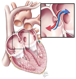
Foetal echocardiograms are used to help identify heart defects before a child is born. The bubble test involves injecting a solution in the blood stream and view the blood flow with ultrasound echo. The bubble study offers a modern technique to test the heart for any abnormalities in terms of an opening in the upper cardiac chambers which can sometimes cause heart failure.
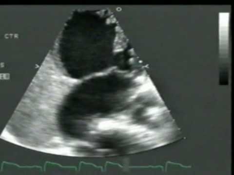
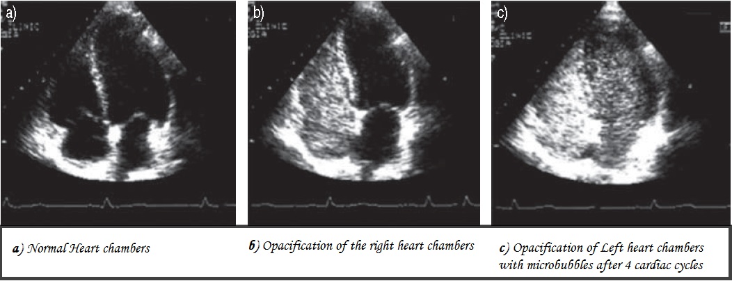
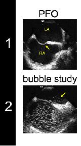
If there is a hole in the heart the bubbles will be visible going from between the ventricles in an unhealthy fashion. There are several types of echocardiograms. This provides a clearer image of the heart because the sound waves do not have to pass through skin muscle or bone tissue.
Your doctor may ask to have a bubble study when the echocardiogram test. During your echo youll be able to see a picture of your beating heart. 8262019 A bubble study gives added information as it can identify potential blood flow issues inside your heart.
If the bubbles are seen on the left side it shows that there is an opening between the two sides of the heart which is abnormal. Echocardiogram with bubble study. During certain portions of the imaging saline with bubbles is.
This fluid then circulates up to the right side of your heart and shows up. It is typically used in conjunction with an echocardiogram in which case doctors often call it contrast echocardiography or a transcranial Doppler study TCD. The sound waves make moving pictures of your heart so your doctor can get a.
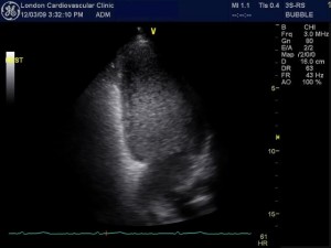
2142021 A bubble echocardiogram is a procedure which is designed to give a doctor an idea of how well someones heart is functioning. The image shown here is enhanced by 3D technology. TCD Bubble Test For Cardiac Shunt Detection PFO Test.
A saltwater solution called saline is mixed with a small amount of air to create tiny bubbles and then injected into your vein. The abnormality can be an atrial-septal defect or a ventricular septal defect. 3302020 Because there are many types of cardiac disease each affecting different parts and functions of the cardiovascular system we need a battery of heart tests to make an accurate diagnosisSome tests such as an echocardiogram are used to identify abnormalities within the heart or blood vessels.
The bubble study helps in finding any hole present in the chambers of the heart and also to find if blood is present inside it causing any trouble. Often the test is to view whether any of the soultion is shunted from the right side through a hole in the wall to the left side. Vascular CenterTCD BUBBLE TEST for Cardiac Shunt Detection PFO TESTZsolt Garami MD.
In a normal heart the bubbles are filtered by the lungs and are seen only on the right side of the heart.
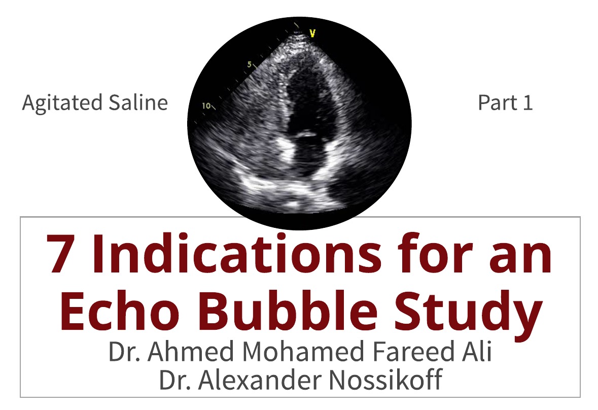 7 Indications For An Echo Bubble Study
 7 Indications For An Echo Bubble Study
 Agitation Saline Injection Test On Contrast Echo Conducted On Rt Arm Download Scientific Diagram
 A Bubble Study To Diagnose Patent Foramen Ovale Youtube
Agitated Saline Bubble Study Youtube
 What Is A Bubble Study Harvard Health
 2d Echo Bubble Study Youtube
 Atrial Septal Defect Wikipedia
 Bubble Contrast Echocardiography Bubble Echocardiogram Test Llc
 Tcd Bubble Test For Cardiac Shunt Detection Pfo Test Zsolt Garami Md Youtube
 Bubble Test Respiratory Diseases
 What Is A Bubble Study How To Effective Bubble Study
Comments
Post a Comment