While only one hip may be imaged your doctor. 7272020 You will have an X-ray before being discharged to make sure that your hip replacement looks normal.
 Hip Replacement With Constrained Liner Radiology Case Radiopaedia Org
Osteolysis or bone loss after total joint replacement knee or hip can be a problem.
Hip replacement x rays. Here the prosthetic hip joint dark left can be seen with the peg implanted in the femur centre-left and. Coloured X-ray of a section through the pelvic the region of a 66-year-old male patient after total hip replacement surgery. Frontal image of the inferior parts of the hip bone to include the ischium bones for measurements.
Images Joey McLeisterStar Tribune MinneapolisMnFriAug. After surgery you will be moved to the recovery room where you will remain for several hours while your recovery from anesthesia is monitored. Aspiration of the hip is the best test for excluding infection.
This gives your doctor a good view of the joint from different angles. Here the prosthetic hip joint dark left can be seen with the peg implanted in the femur centre-left and. You will usually have two X-rays taken of your hip one from the front and one from the side.
The images suggest this problem didnt just happen but was developing over time. 9132019 Post-operative projectional radiography X-ray is routinely performed to ensure proper configuration of hip prostheses. X-rays are carried out at Circle by a diagnostic radiographer.
A standard hip X-ray examination generally includes an anteroposterior PA image and a lateral image. 11122018 X-rays are complementatry to other studies. Such joint replacement orthopaedic surgery is generally conducted to relieve arthritis pain or in some hip fractures.
As Ive blogged before there are many things that cause hip pain such as the SI joint so regrettably this may mean that we have a reasonable number of patients undergoing an invasive and risky hip replacement who dont need the surgery. 27 2004--Marjorie Therres X-ray shows her two hip replacements. Hip replacement surgery can be performed as a total replacement or a hemi half replacement.
In this case non-cemented components were used. Known as the antero-posterior AP view this is taken from the front of your hip. Coloured X-ray of a section through the pelvic the region of a 66-year-old male patient after total hip replacement surgery.
How is a hip X-ray done. Most partial hip replacement surgeries can be performed in less than one hour in a hospital or surgical center. Hip replacement surgery is a procedure in which a surgeon makes an incision over the side of the thigh removes the diseased parts of the hip joint and replaces them with new artificial parts.
Hip replacement is a surgical procedure in which the hip joint is replaced by a prosthetic implant that is a hip prosthesis. The hip joint can be imaged under various angles. The pieces may be made of metal plastic ceramic or a combination of these materials.
Frontal and axial image of the hip joint. THR is a surgical procedure that replaces the diseased cartilage and joint with artificial materials made of metal and plastic. These parts mimic the way a normal hip joint works.
With the aid of X-rays CT scans andor MRIs the surgeon diagnoses a condition known as osteolysis. The patient is placed under general anesthesia and the front of the leg and hip are sterilized. Total hip replacement THR is a process in which the hip joints are replaced with artificial joints or prosthesis.
DePuy Synthes Revision Solutions. X-ray of a hip replacement in a 50 year old woman - hip replacement xray stock pictures royalty-free photos. X-rays before and after total hip replacement.
1242021 X-rays will be taken to confirm that the hip replacement procedure was a success. 9262013 This study suggests that a hip x-ray is sorely lacking in any ability to determine who needs a hip replacement. Recovering from a hip replacement Staff on the ward will look after you when you come back from the operating theatre and you will usually be able to have something to drink within an hour or so after you get back to the ward.
In-111 WBC and Tc-99m sulfur colloid scan is. The hip X-ray is used primarily to demonstrateexclude a fracture. Hip X-rays are also frequently opted for as initial test in chronic hip symptoms eg.
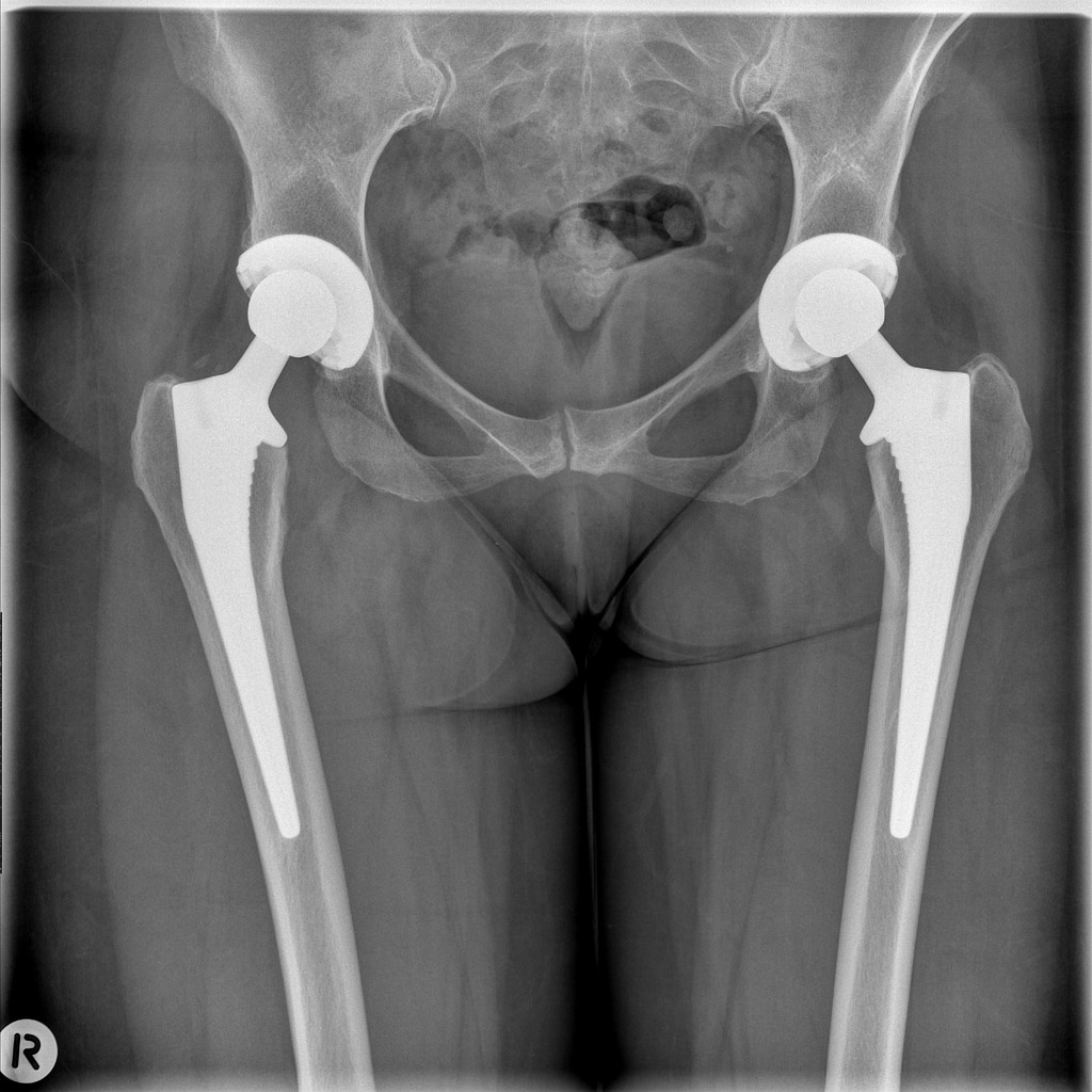
Normal Pelvis And Both Hips With Bilateral Total Hip Replacements Radiology Case Radiopaedia Org
 Main Complications Of Hip Arthroplasty Pictorial Essay
 Post Charnley Left Total Hip Replacement Plain X Ray Of The Pelvis Download Scientific Diagram
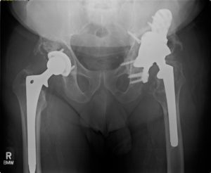 Revision Total Hip Replacement Richmond Va Dr Wind Orthova
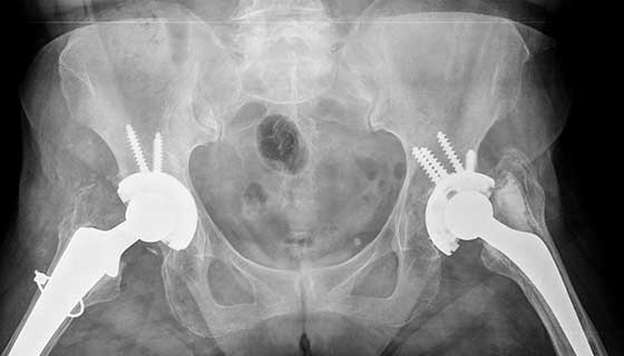
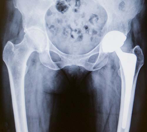
 Hip Replacement Periacetabular Osteolysis Radiology Case Radiopaedia Org

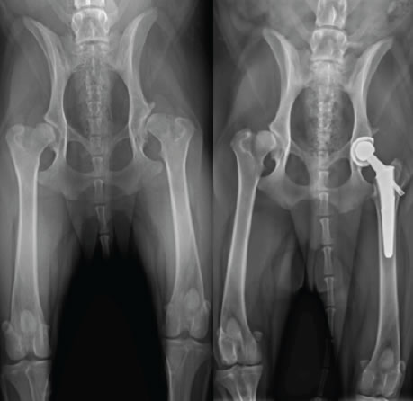
 File X Ray Of Hips With A Hemiarthroplasty Jpg Wikimedia Commons
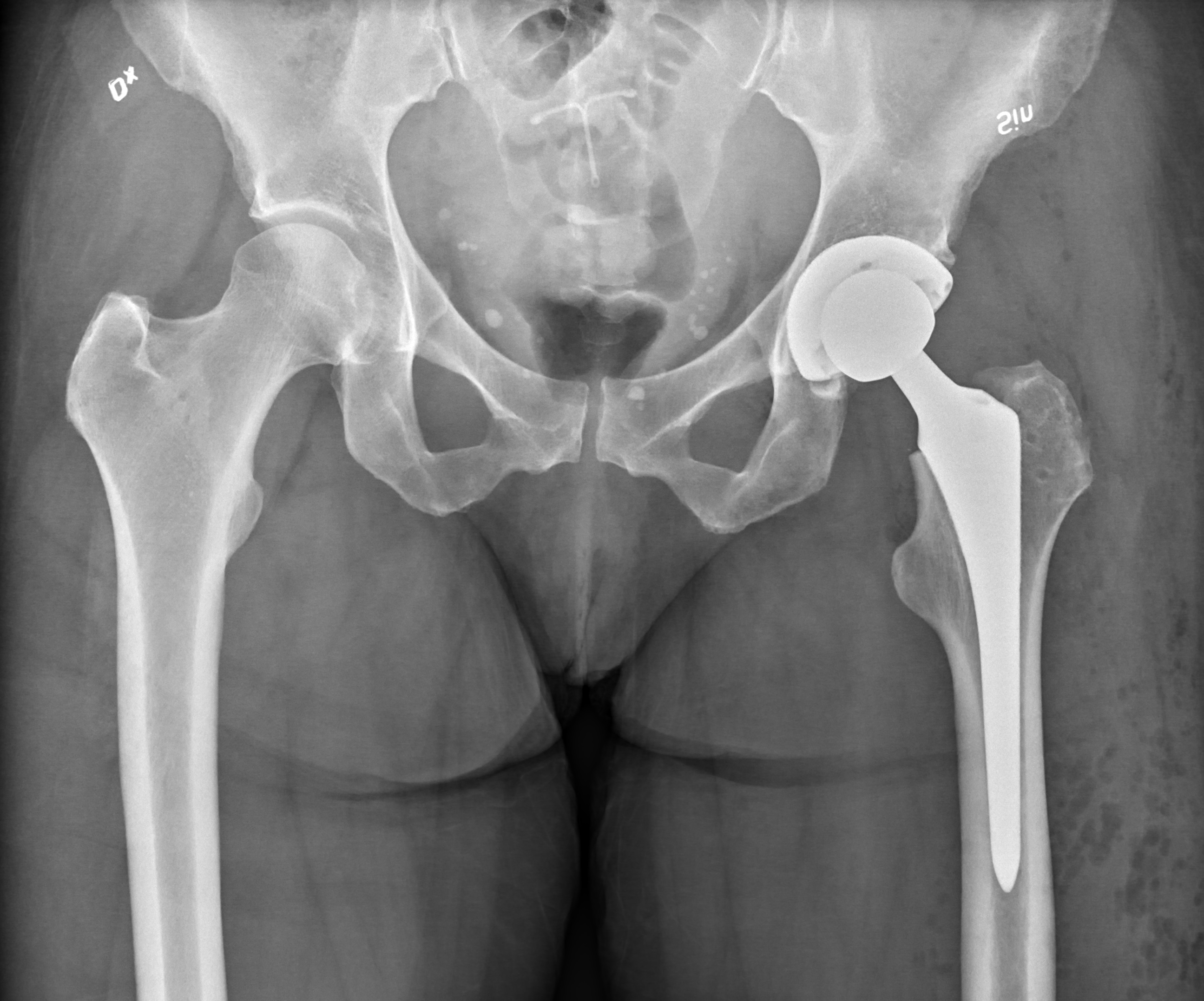
 X Ray Pelvis Following Total Hip Replacement Download Scientific Diagram
 Hip Replacement Wikipedia
 Hip Replacement Surgery Procedure Types And Risks Hss
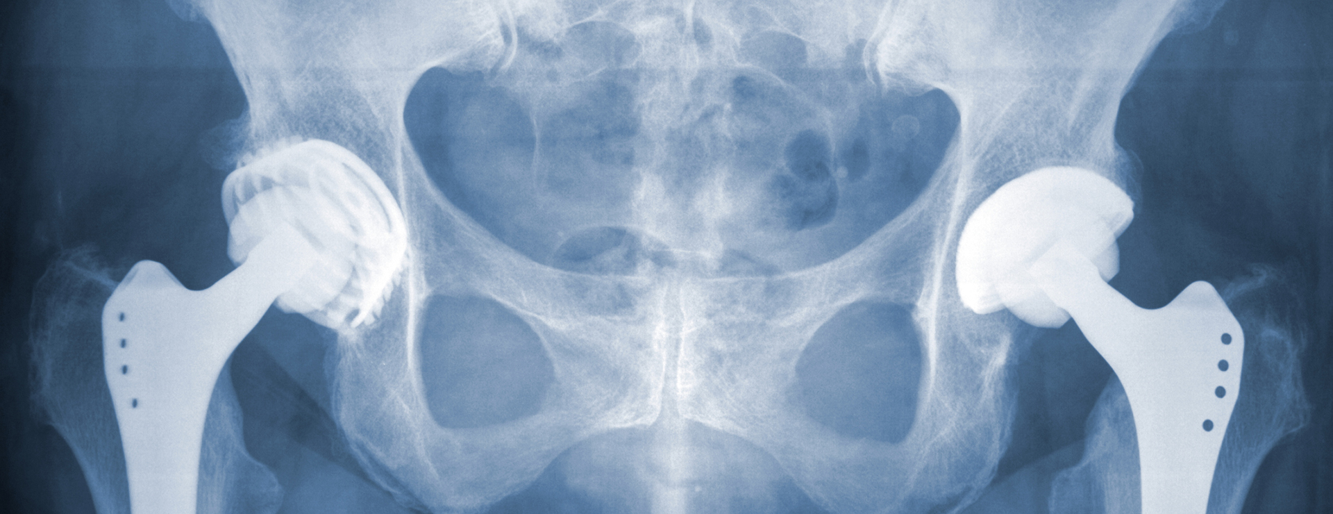
 X Ray Scan Image Of Hip Joints With Orthopedic Hip Joint Replacement Or Total Hip Prosthesis On Right Side Implant Head And Screws In Human Skeleton In Blue Gray Tones Movement Orthopedics
Comments
Post a Comment