Interestingly there have been no published articles with current data considering the. 112009 A straight lateral view for an asymmetry seen only on a mediolateral oblique MLO view and a rolled view for an asymmetry seen only on a craniocaudal CC view should be obtained.
 X Ray Digital Mammogram Or Mammography Image Mlo View Stock Image Image Of Bilateral Diagnostics 168025041
The technologists alertness and diligence are the keys to good positioning.

Mlo view mammogram. Achieving an optimal mammographic image requires full patient cooperation. 7142016 There is a mediolateral oblique MLO view which is looking through your breast from the side. This is why it was chosen for DBT.
Standard Digital Mammogram Images. Ideal MLO positioning should permit the breast to be imaged from high in the axilla down to and including the IMF. The following sections describe these views.
More specialized views are described in the ACR Mammography Quality Control Manual ACR 1999. A technically adequate exam has the nipple in profile allows visualization of the inframammary fold and includes the pectoralis muscle extending down to the posterior nipple line an oblique line drawn straight. The MLO view allows visualization of the largest amount of breast tissue.
If no oblique image is taken this mediolateral angle may be preferable to the latero-medial view LM. The views are usually used for all routine screening clients. 552019 An asymmetrical density mammogram in terms of the first mammographic finding usually refers to an opacity obscured view in part of the breast which is visible on only one projection or one view or angle of the X-ray.
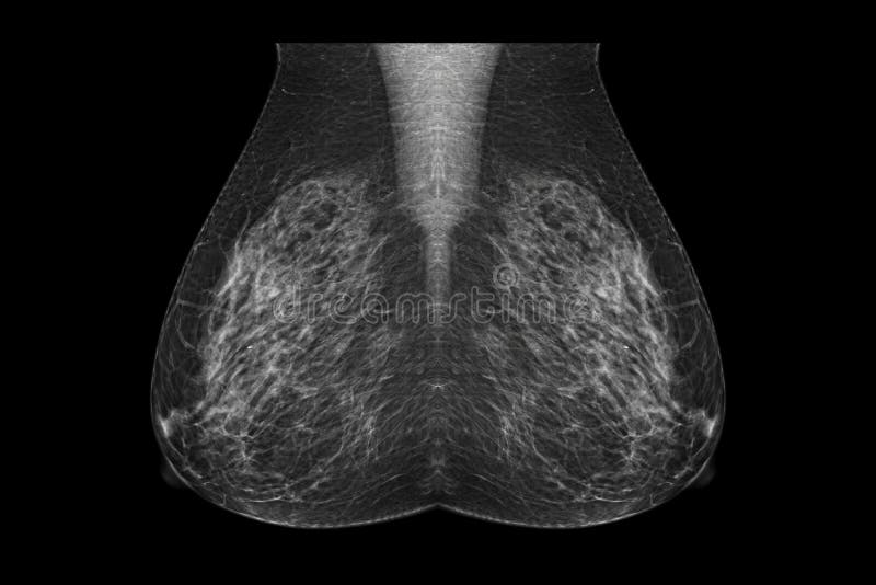
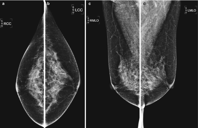
It is the most important projection as it allows depiction of most breast tissue. Incomplete - Additional imaging evaluation andor comparison to prior mammograms is needed. Standard views are bilateral craniocaudal CC and mediolateral oblique MLO views which comprise routine screening mammography.
6112017 Two view mammography combining the MLO and the CC projections creates a three dimensional representation of the breast. Craniocaudal CC left and mediolateral oblique MLO right views of 48 year-old woman with palpable lump in upper outer left breast show heterogeneously dense tissue which may obscure small masses. Two projections are the basic components of the standard routine mammogram.
Mammogram MLO view shows deodoranttalcum powder artifact mimicking microcalcifications in the axilla. This allows for maximum visualization of the breast tissues while obtaining the information required in order to gain a three-dimensional understanding of the visualized structures. The MLO provides the greatest amount of coverage for a single projection.
A screening mammogram is composed of a CC and MLO view of each breast. Radiologists prefer the MLO view or from the side-at an angle view to a. Patient-related artifacts positioning as well as motion artifacts occur more frequently in.
Two view mammography is described for both initial and incident or subsequent screening rounds by the UK BreastScreening Programme American College of Radiology BreastScreen Australia and BreastScreen Aotearoa. This means the radiologist may have seen a possible abnormality but it was not clear and you will need more tests such as another mammogram with the use of spot compression applying compression to a smaller area when doing the mammogram. MLO Views Supplemental views Advanced Health Education Center Objectives To understand the role of additional mammographic views Objectives To view diagnostic mammography as a tailored examination To select appropriate diagnostic views By the radiologist By the technologist Clinical Correlation Information from the patient.
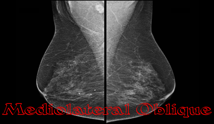
Mediolateral oblique MLO visualization of the inframammary fold IMF was obtainable only 49 of the time. In addition there is a craniocaudal CC view which is looking through your breast from above. The mediolateral oblique MLO view is one of the standard views obtained during every screening exam.
That is unless there is a contraindication screening mammograms consist of these 4 views. The Mediolateral View also called the medio-lateral view or ML is taken from the center of the chest between the breasts outward. The single most useful mammographic projection of the breast is the MLO image.
Screening mammograms should only be performed on women that do not have an area of concern. Not all 4 views are always performed in all mammogram studies. The mediolateral oblique MLO view is one of the two standard mammographic views alongside the craniocaudal CC view.
562019 The medio-lateral view ML is a view from the center of the chest outward whilst the latero-medial view or LM is a view from the outer side of the breast towards the middle of the chest. Typically the radiologist will request additional views from other X-ray angles as an immediate follow up. A thorough review of proper positioning of the craniocaudal and mediolateral oblique views for the screening mammography patient will be discussed in detail.
412021 The routine two-view mammogram consists of a CC projection and a MLO projection ACR 1993. If the asymmetry is maintained even after the angle of projection is changed additional views in other projections should be obtained. The craniocaudal CC and the mediolateral oblique MLO views Fig.
This outcome was largely due to variations in body habitus and other patient issues. Although mammography patient positioning is the one element that is practiced most frequently it is the one criteria that is shown to be most deficient. The patients cancer in the area marked by red arrows is not well seen.
 9 Mammogram A Mlo View B Cc View Download Scientific Diagram
 Pectoralis Nipple Line On A Cc View And B Mlo View Download Scientific Diagram
Mediolateral Oblique View Radiology Reference Article Radiopaedia Org
 Digital Mammogram Both Image Photo Free Trial Bigstock
 X Ray Digital Mammogram Or Mammography Image Mlo View Stock Photo Image Of Breast Illness 168025116
 A Cc View B Mlo View Cc Cranial Caudal Mlo Download Scientific Diagram
 Right Mammogram Mlo View 25 Mm Lobulated Mass Lesion Surrounded By Download Scientific Diagram
![]() Shutterstock Puzzlepix
Shutterstock Puzzlepix
 Mammography Techniques Mammoguide Learn Breast Imaging
 The First Question Radiology Key
 Mammography Techniques Positioning And Optimizing Image Quality Oncohema Key
 Typical Mammogram In Mlo View From Clinical Dataset In The Roi Download Scientific Diagram
 Using The Location Information From Mlo As Well As Cc View A Marker Download Scientific Diagram
 Pin On Misc
Comments
Post a Comment